0146-00 Internal Organs
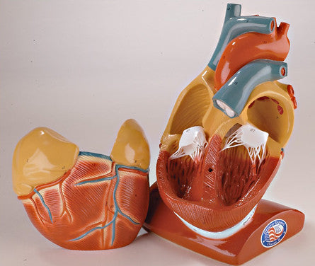
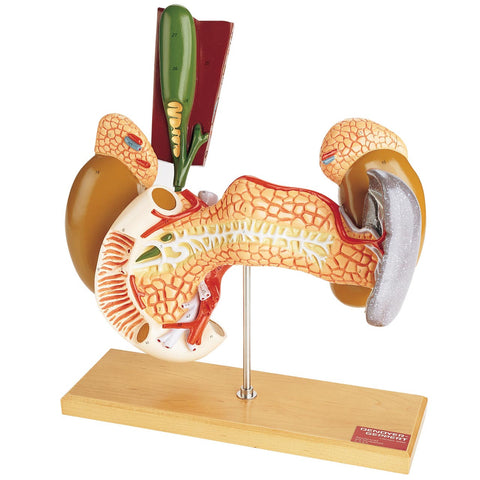
On this model the spleen is shown drawn away from the left kidney to expose its renal surface. The peritoneum is removed except over the spleen. The pancreas is shown somewhat lower than normal.
-
Body of pancreas
-
Head of pancreas
-
Tail of pancreas
-
Pancreatic duct (Duct of Wirsung)
-
Accessory pancreatic duct (Duct of Santorini)
-
Papilla major of duodenum
-
Papilla minor of duodenum
-
Duodenum
-
Descending portion of duodenum
-
Inferior portion of duodenum
-
Ascending portion of duodenum
-
Right suprarenal gland
-
Left suprarenal gland
-
Right kidney
-
Left kidney
-
Spleen, drawn away from the left kidney to
expose the renal surface
-
Hilus of spleen
-
Renal surface
-
Gastric surface
-
Diaphragmatic surface
-
Anterior margin
-
Upper extremity
-
Lower extremity
-
Gastrolienal ligament
-
Portion of inferior surface of liver
-
Gallbladder
-
Fundus of gallbladder
-
Neck of gallbladder
-
Cystic duct with spiral valve
30. Hepatic duct
31. Common bile duct
32. Coeliac artery
33. Gastroduodenal artery
34. Superior pancreatico-duodenal artery 35. Hepatic artery
36. Superior mesenteric artery
37. Intestinal arteries
38. Middle colic artery
39. Splenic artery
40. Suprarenal arteries and veins
41. Portal vein
42. Superior mesenteric vein
43. Intestinal veins
44. Middle colic vein
45. Splenic vein
46. Main trunk of portal vein
47. Left gastric artery
48. Splenic artery
49. Right renal artery
50. Right renal vein
51. Left renal artery
52. Left renal vein
53. Hilus of kidney
54. Right ureter
55. Left ureter
56. Branches of portal vein within liver 57. Branches of hepatic veins within liver 58. Branches of hepatic ducts within liver 59. Branches of hepatic artery within liver
We Also Recommend


