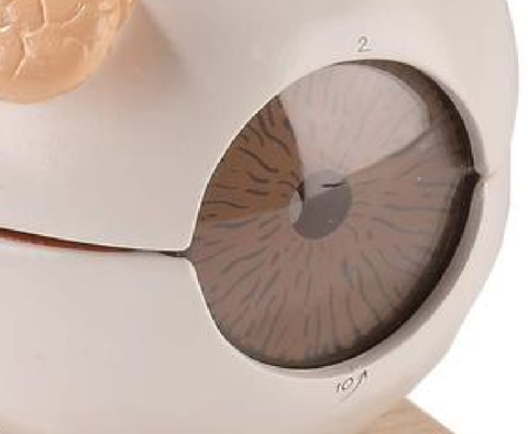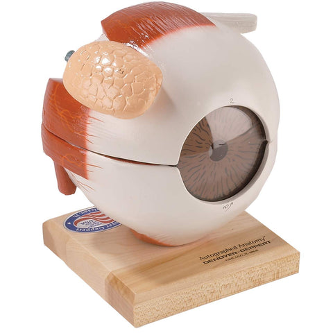
0102-00 Giant Five-part Eye Model
0102-00 Giant Five-part Eyeball
Six-times life-size, this unbreakable vinyl plastic replica is packed with useful teaching features.
On the exterior of the eyeball is the cornea, through which the iris and pupil are visible, the large lacrimal (tear) gland, attachments for all six extrinsic muscles of the eye, the optic nerve roots, and surrounding blood vessels.
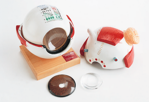
The eyeball divides in half for study of the interior including the meticulously hand-painted choroid coat of the retina. Here, several removable parts are discovered such as the vitreous ball, iris/cornea, and Lucite lens, capable of magnifying and inverting images when removed from the model. Rods, cones, and other retinal microstructures are detailed in a highly-magnified, diagrammatic cross-section. 42 hand-coded features are identified in the corresponding key.
-
Superior rectus muscle
-
Sclera
-
Retina
-
Choroid layer
-
Vitreous body
-
Conjunctiva*
-
Ciliary body
-
Suspensory ligament*
-
Iris*
-
Cornea
-
Aqueous humor*
Lens*
Pupil*
Ciliary muscle
Inferior rectus muscle
Location of optic nerve exit and blind spot Yellow spot with fovea centralis retinae Internal rectus muscle
External rectus muscle
Tendon of superior oblique muscle Inferior oblique muscle
Lacrimal gland
-
Pigmented layer
-
Layer of rods and cones
-
External limiting membranes
-
Outer nuclear layer
-
Outer plexiform layer
-
Inner nuclear layer
-
Inner plexiform layer
-
Ganglionic layer or layer of nerve cells
-
Stratum opticum or layer of nerve fibers
k. Internal limiting membrane l. Rods
m. Rod granules
n. Cones
o. Cone granules
p. Horizontal cells
q. Bipolar cells
r. Amacrine cells
s. Sustentacular fibers of Müller t. Ganglionic cells
We Also Recommend